NeuroSculpt: Forecasting Brain Structure 9 Years Ahead Using Structural MRI
NTNU, Trondheim, Norway
June 2024
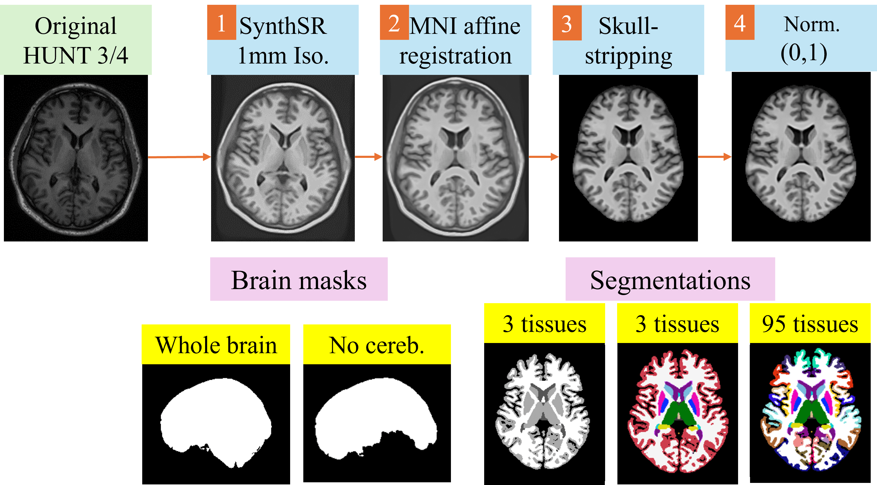
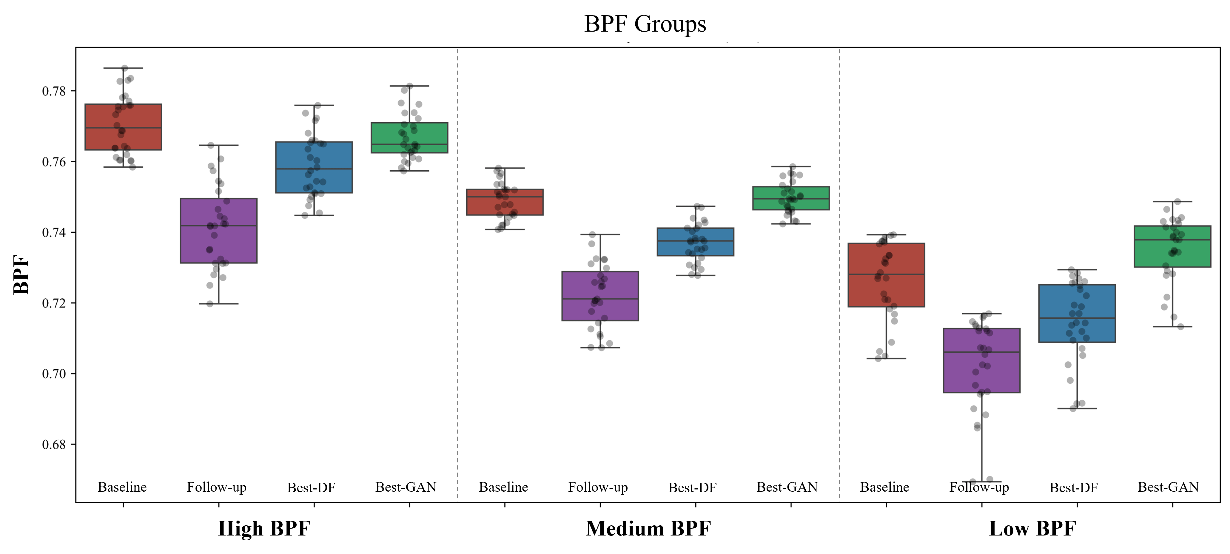
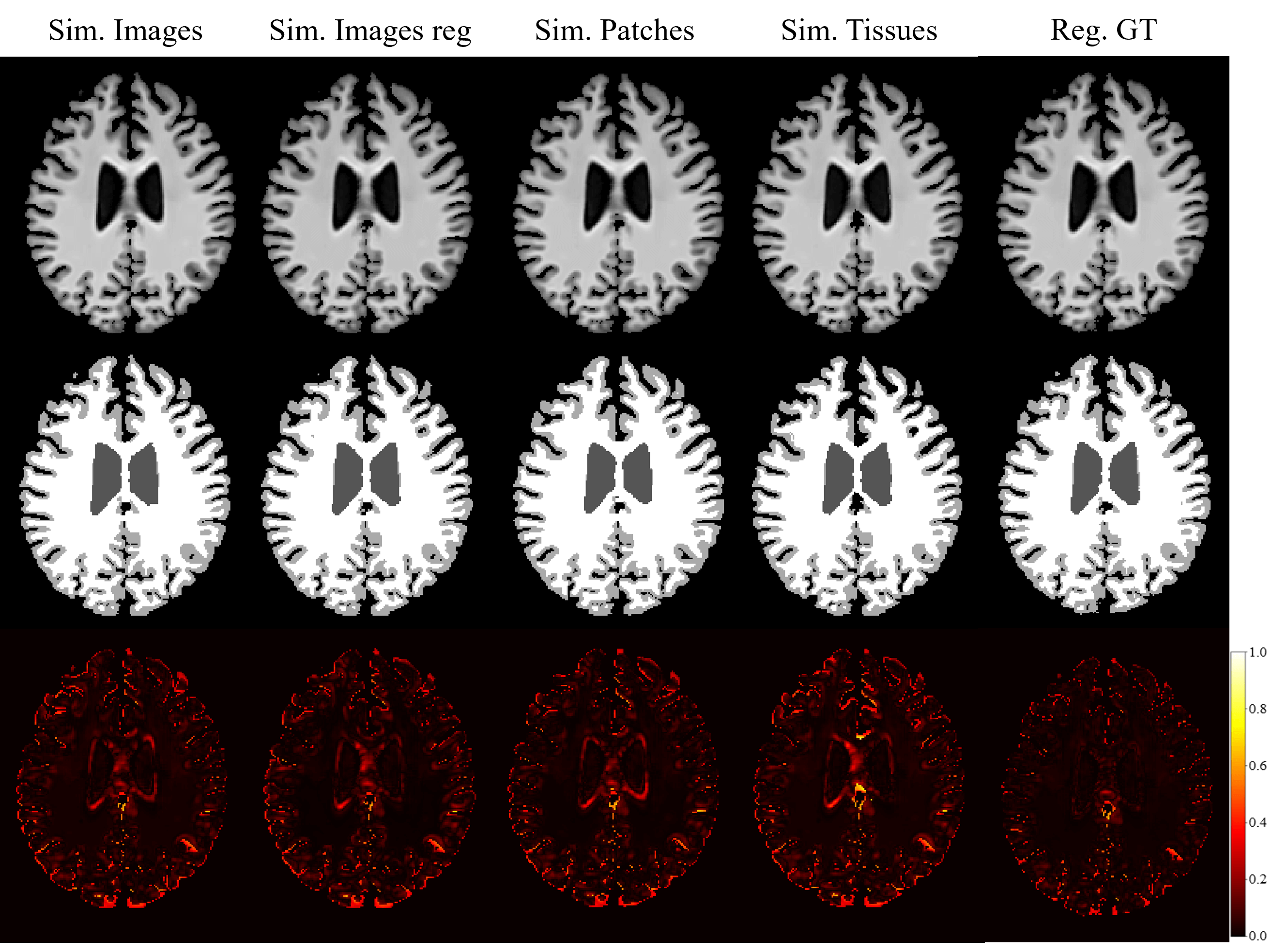
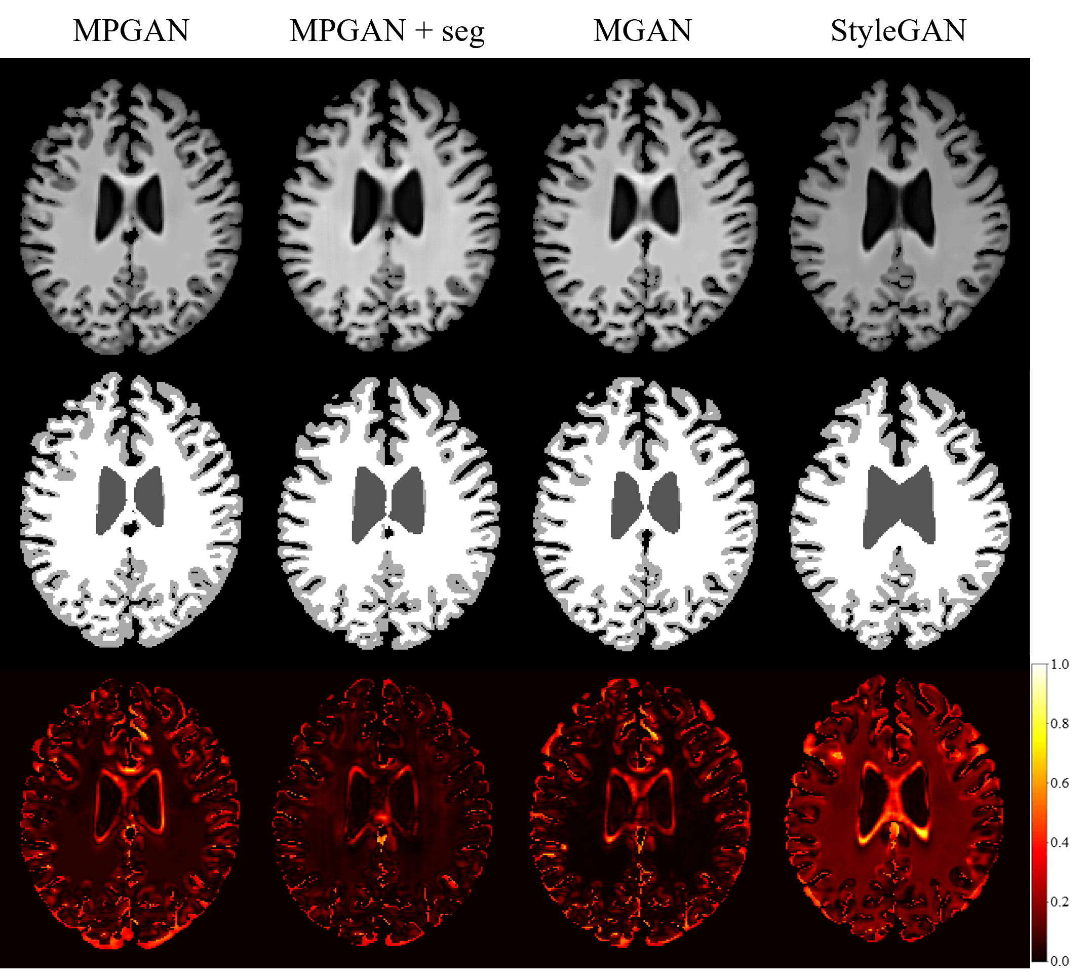
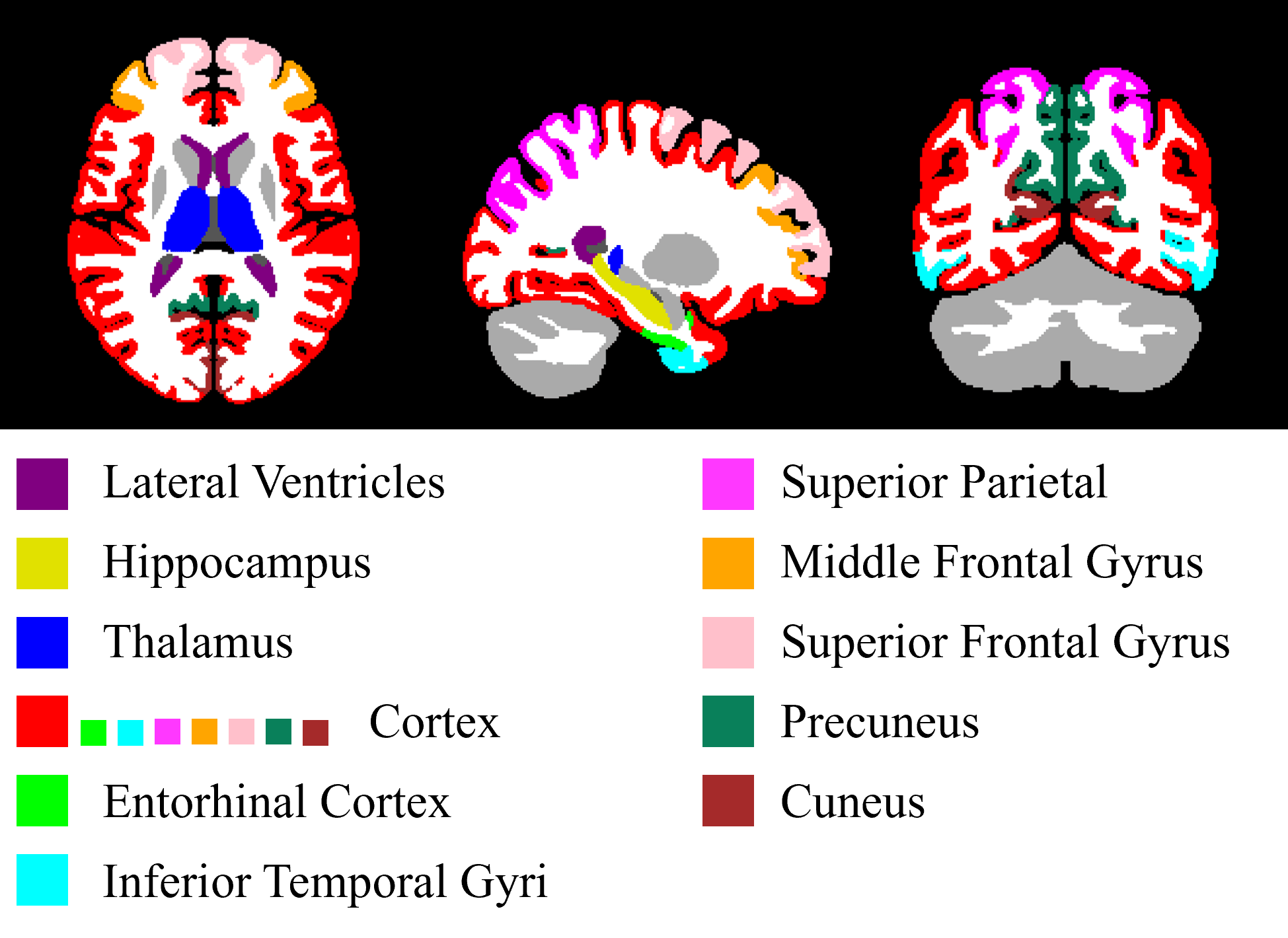
This master thesis focuses on predicting structural brain changes over nine years in a healthy adult population using 3D T1-weighted MR images. Early detection of brain changes is crucial for preventing neurodegenerative diseases like dementia and Parkinson’s. The thesis explores two methods: Deformation Fields (DFs) and deep learning techniques based on Generative Adversarial Networks (GANs). Results showed that DF-based techniques were more effective and stable, capturing subtle changes in the thalamus and cortex, as well as significant changes in the ventricles. In contrast, GANs primarily predicted volumetric changes in the ventricles. This research provides a foundation for future studies, emphasizing the potential of DF-based approaches and suggesting improvements for GAN methods.
Hybrid Atlas and Cascade U-NET Brain Tissue Segmentation
UdG, Girona, Spain
January 2024
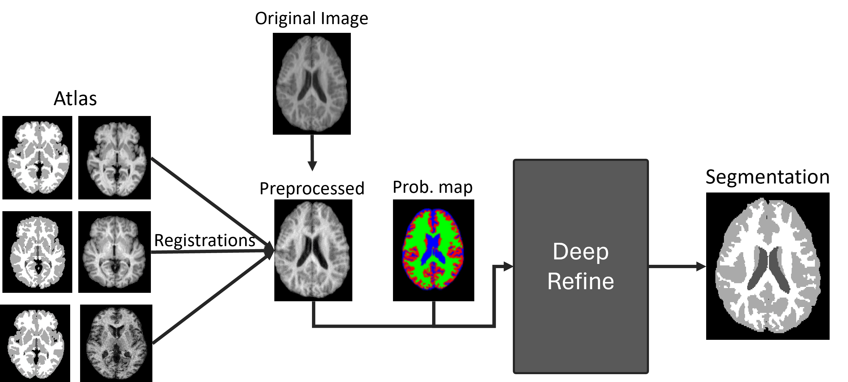
This project aims to develop an AI-based method for segmenting the three main brain tissues: white matter (WM), grey matter (GM), and cerebrospinal fluid (CSF), using magnetic resonance imaging (MRI). The approach integrates a probabilistic atlas as prior spatial information into a deep learning model to achieve accurate segmentation.
Image registration of chest CT volumes
UdG, Girona, Spain
January 2024
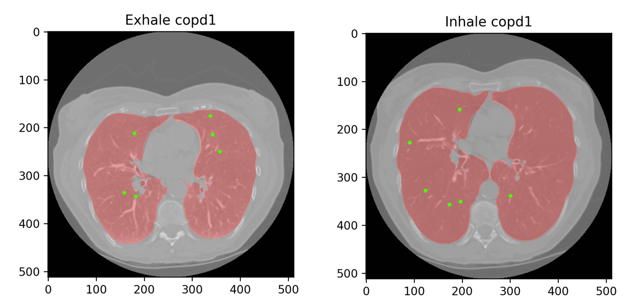
This project compares image registration methods for 4D CT scans of the lungs during inhalation and exhalation. It contrasts Elastix's traditional affine and BSpline approaches with VoxelMorph's deep learning algorithm, achieving an average Target Registration Error of 4.12mm. The study aims to enhance alignment accuracy for Chronic Obstructive Pulmonary Disease diagnosis and treatment.
Dermoscopic diagnosis using Deep Learning
UdG, Girona, Spain
January 2024
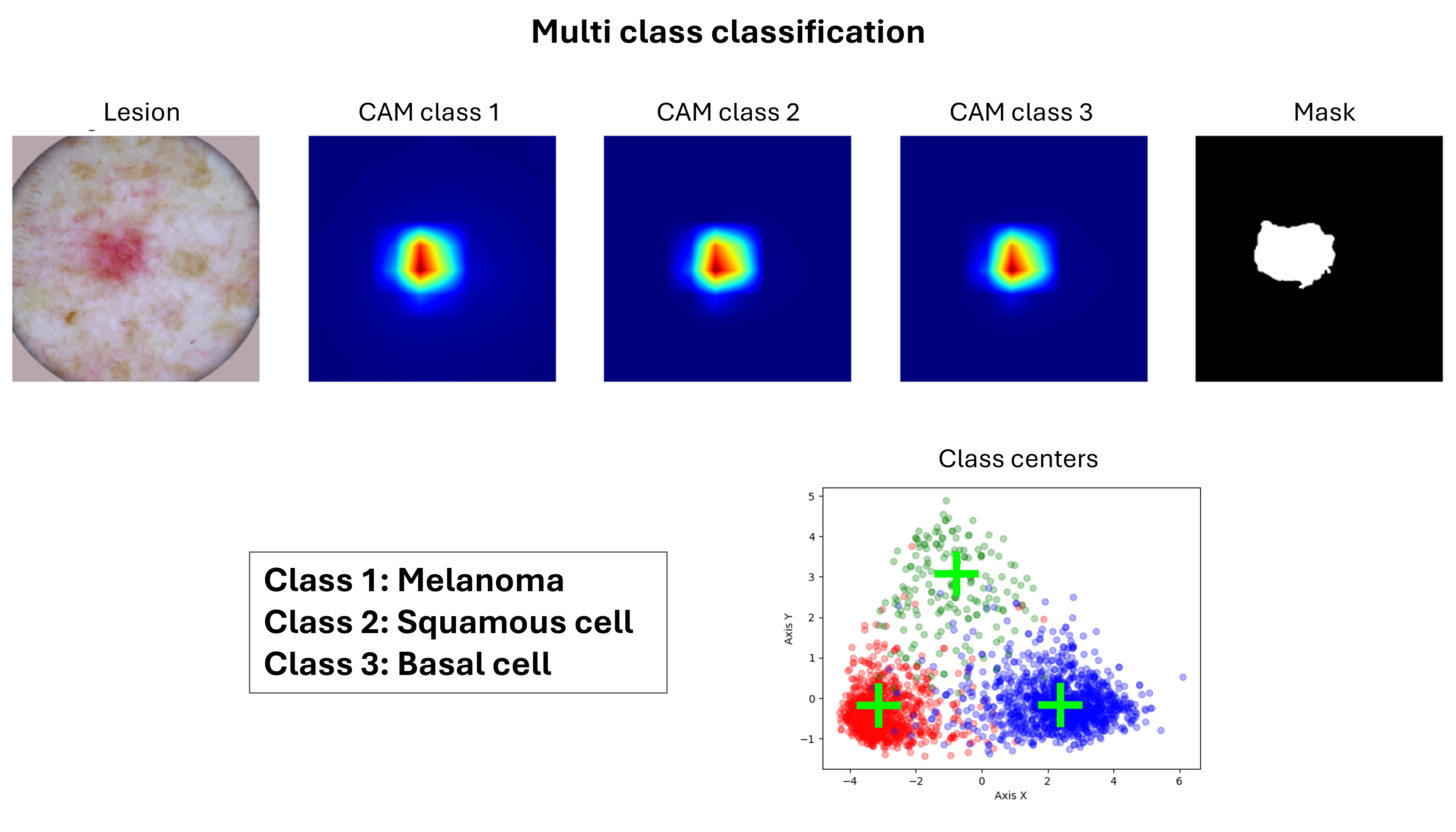
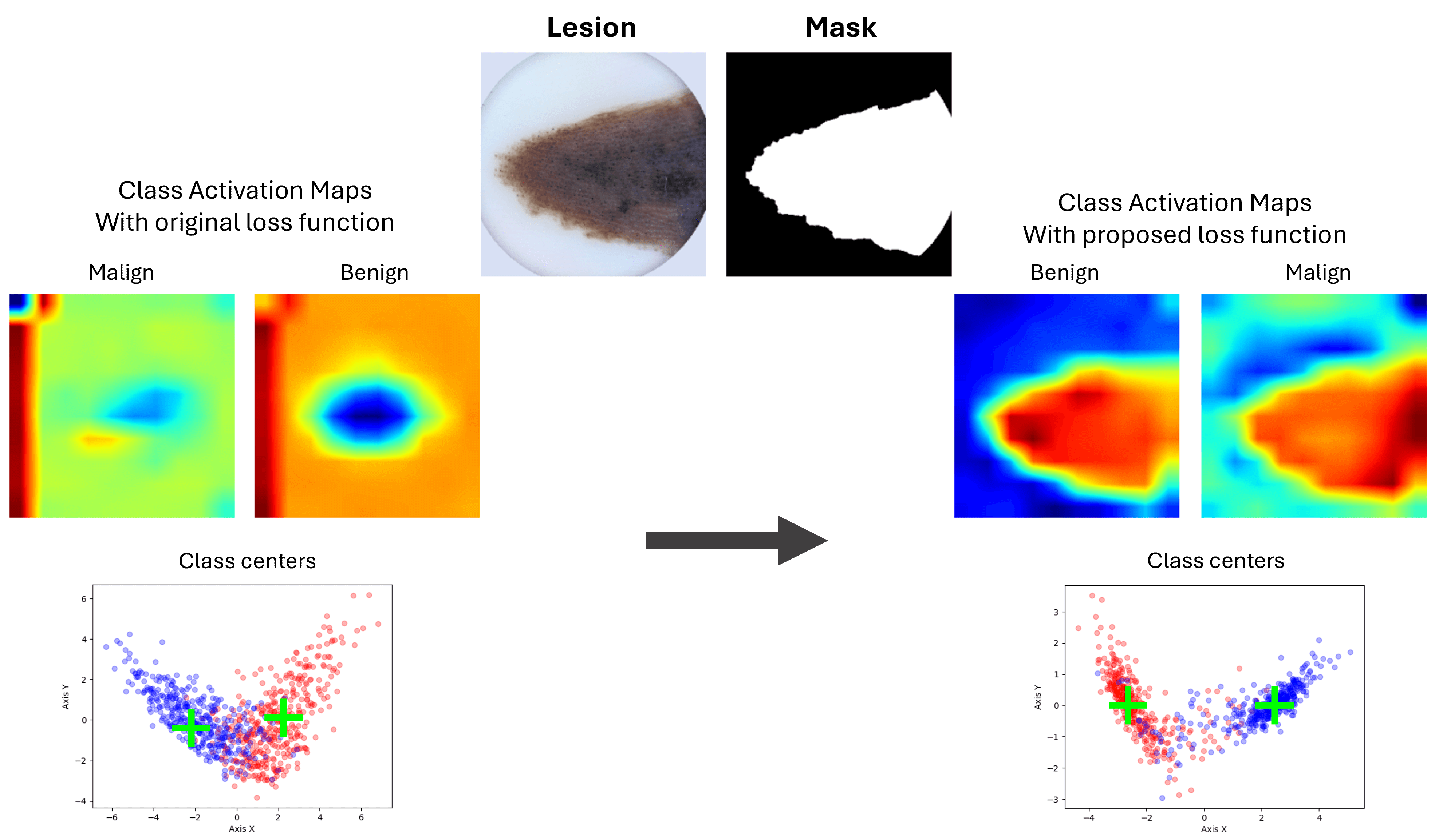
This project focuses on skin lesion detection using deep learning architectures. Two main tasks were addressed: binary classification distinguishing between benign lesions and melanomas, and multi-class classification differentiating between melanoma, basal cell carcinoma, and squamous cell carcinoma. We implemented two innovative loss functions: Mask Loss, based on the Pearson correlation coefficient between the actual mask and the Class Activation Maps (CAMs), and Center Loss, which utilizes the Euclidean distance between image features and class centers. These functions enhance feature discrimination across classes while minimizing intra-class variance.
WoundCareBot
UdG, Girona, Spain
January 2024
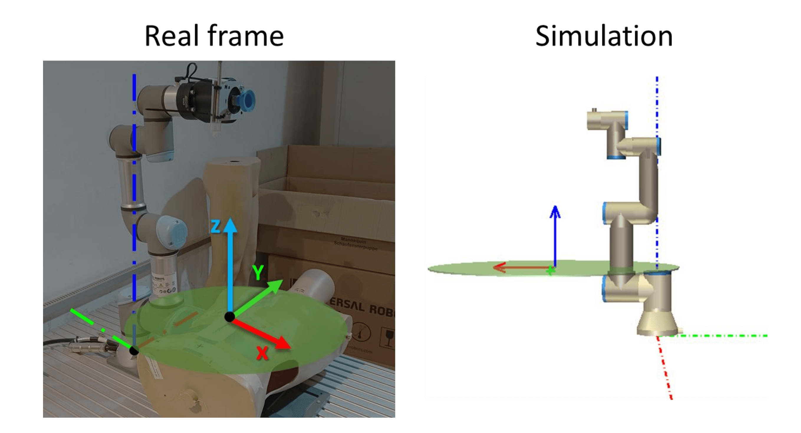
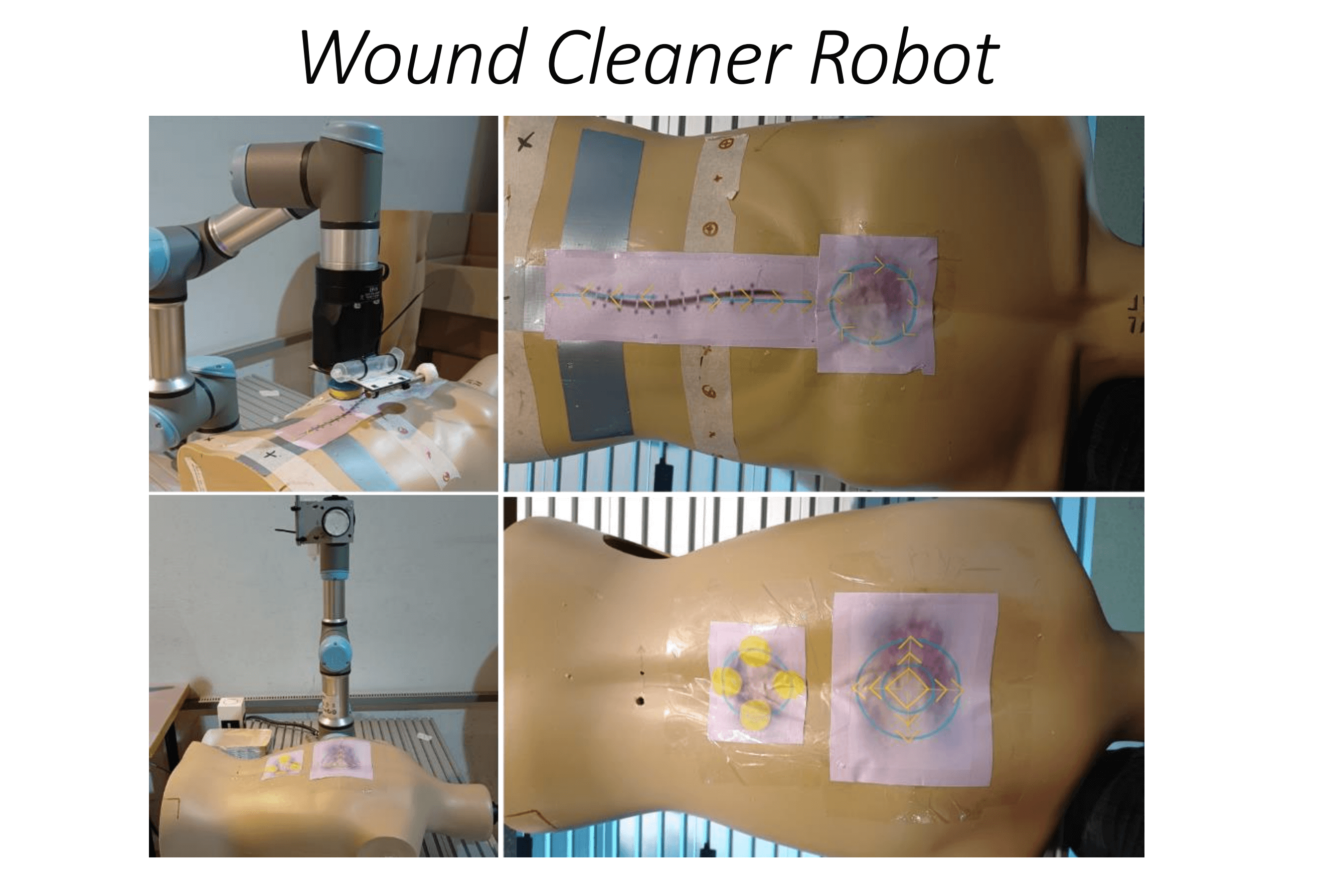
The goal of this project is to develop ‘WoundCareBot’, a robotic system designed to automate precise wound cleaning. Using a Universal Robot with 6 degrees of freedom, the system performs four key movements based on wound type: linear, circular, circular-push, and circular-exterior. These motions are tailored to different wound cleaning needs, such as surgical wounds, burn wounds, and diabetic foot ulcers, ensuring proper force is applied to reduce infection risk and promote healing. WoundCareBot aims to enhance efficiency and precision in wound care, improving patient outcomes in medical settings.
Dermoscopic diagnosis using image analysis techniques
UdG, Girona, Spain
December 2023
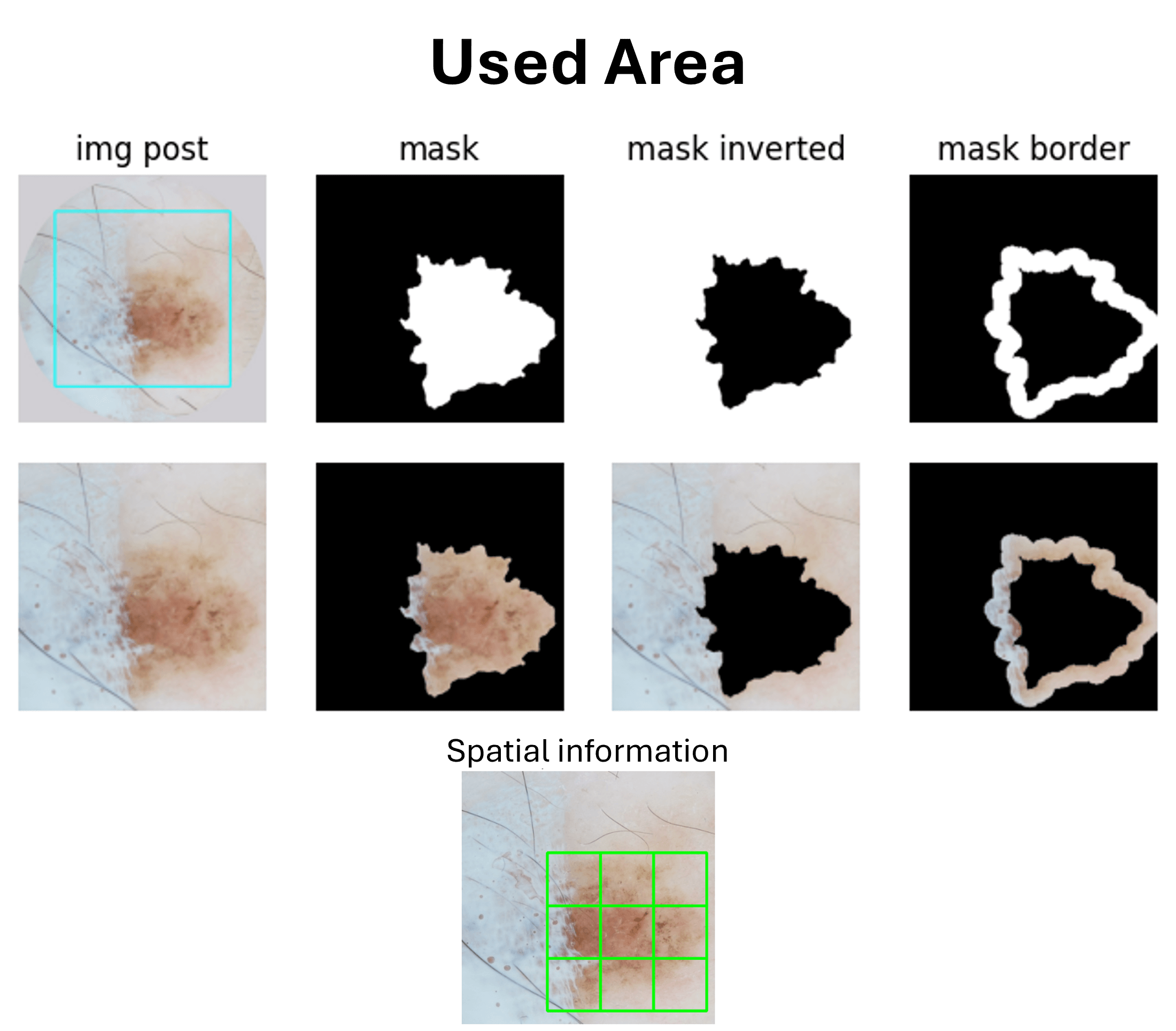
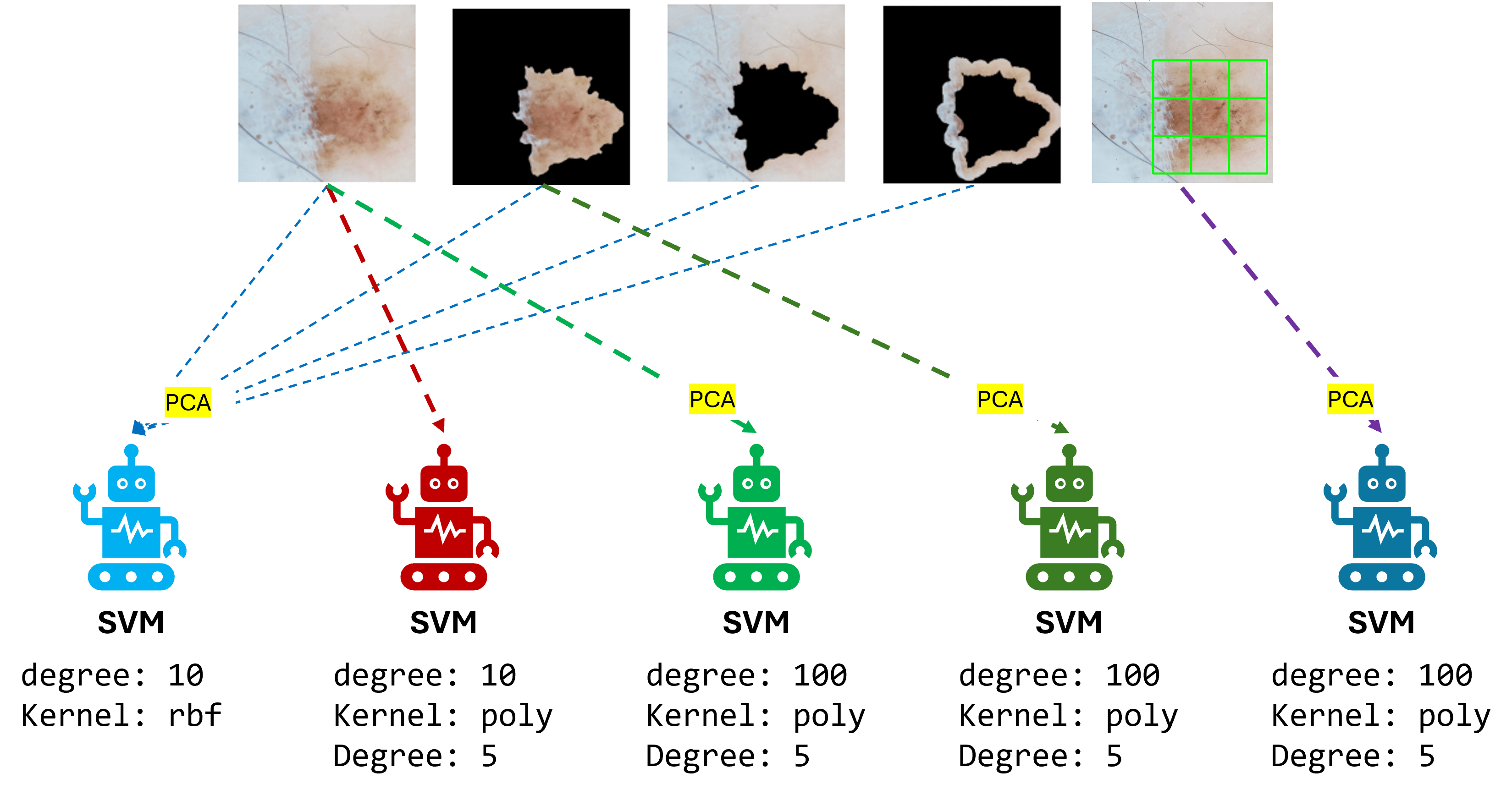
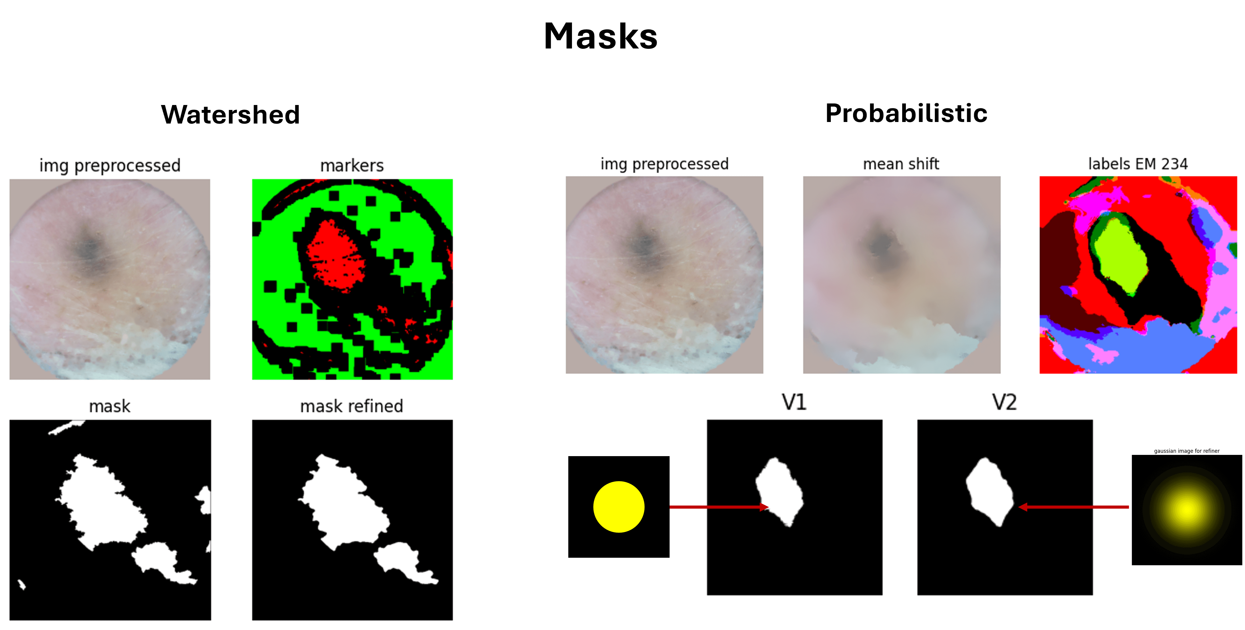
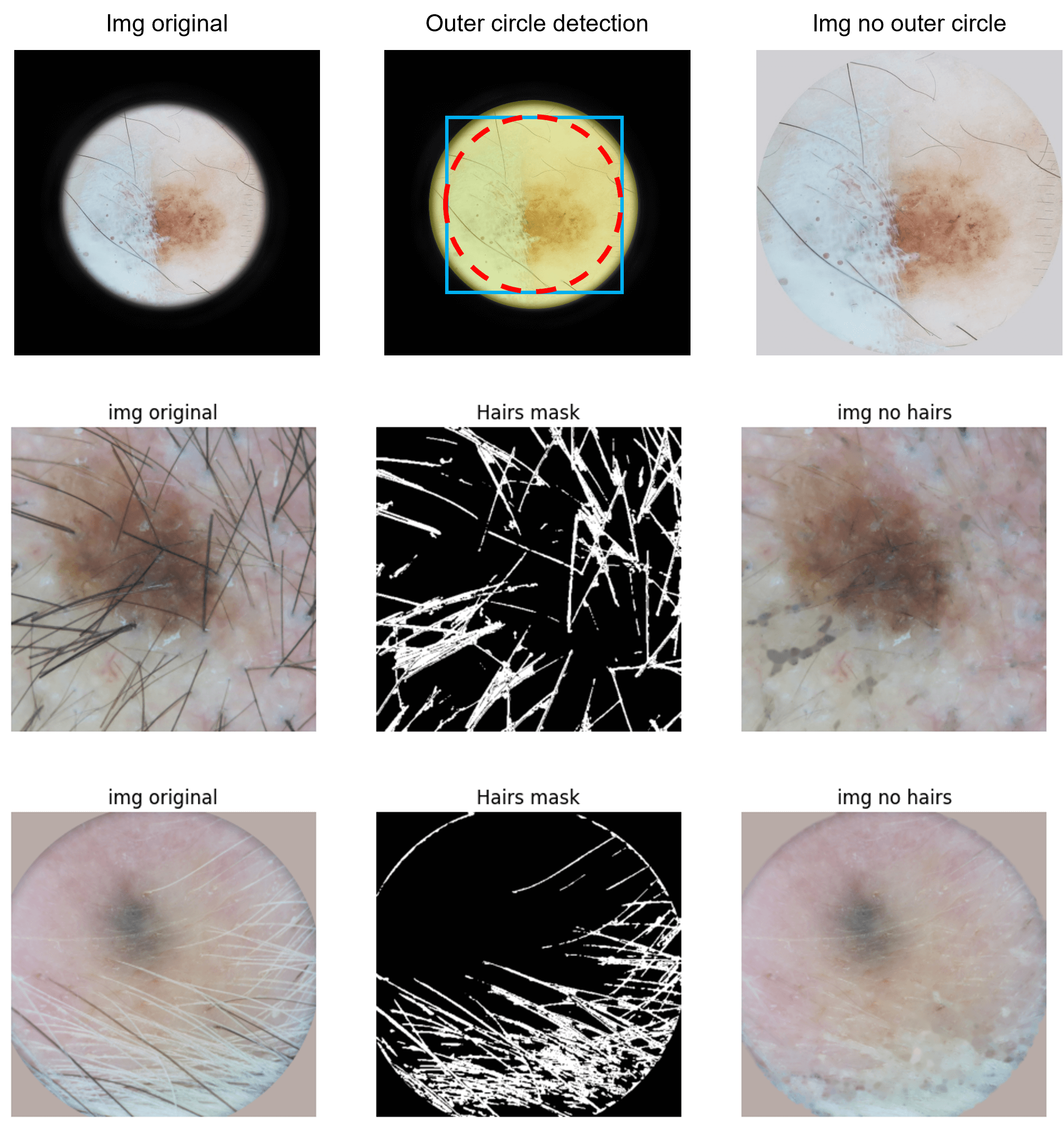
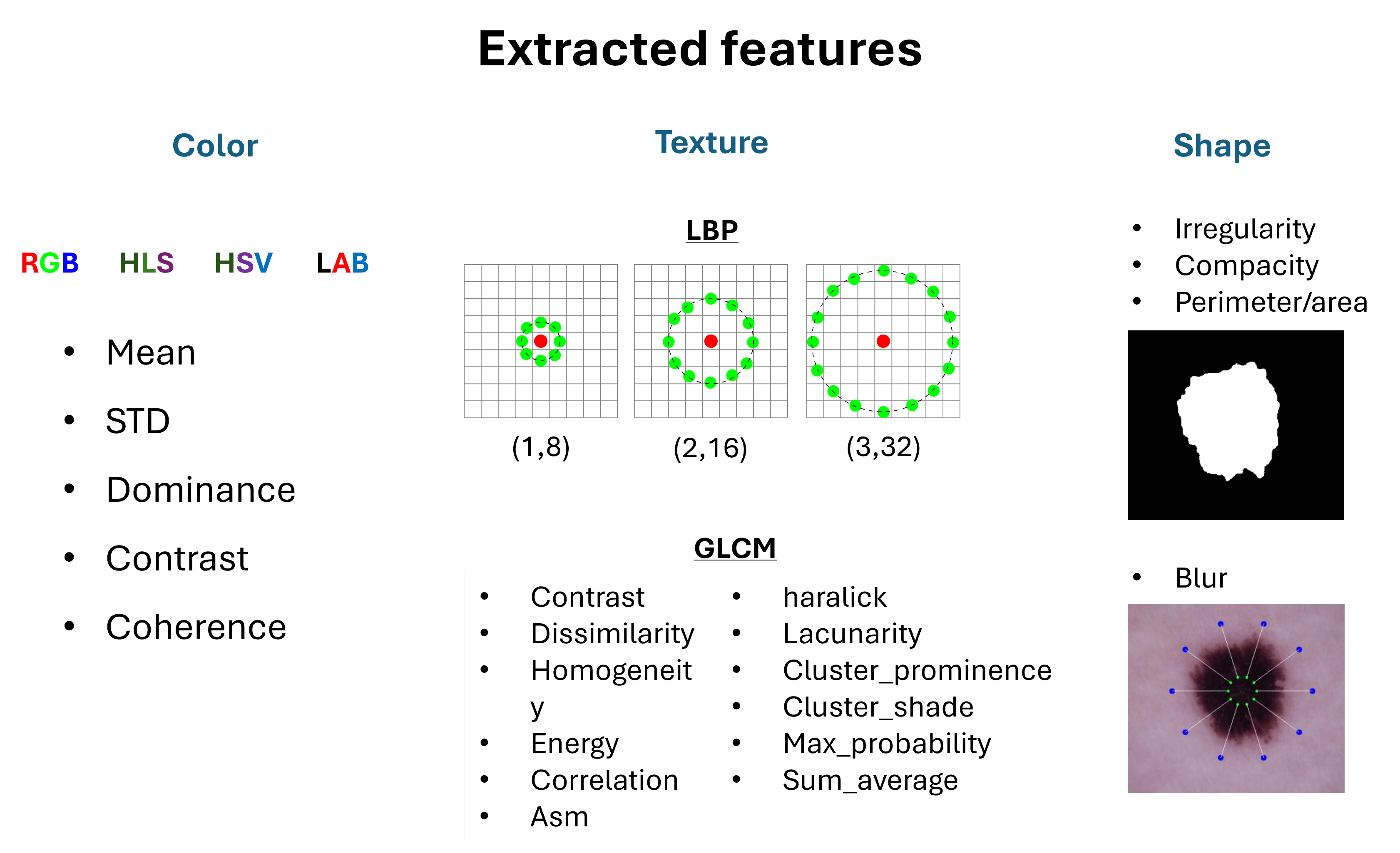
This project aims to develop a computer-aided diagnosis algorithm for skin lesions in dermoscopic images using image analysis techniques. It involves binary classification to distinguish between benign lesions and melanomas, as well as multi-class classification for melanoma, basal cell carcinoma, and squamous cell carcinoma. The algorithm generates probabilistic masks obtained from Gaussian Mixture Models and refines these masks using watershed segmentation to enhance lesion delineation. Various types of features (texture, color, shape) are extracted from different image regions, and five weak SVM classifiers are trained on these features. In the multi-class approach, minority class scores are weighted to address class imbalance, and the classifiers’ outputs are combined using the Dempster-Shafer method, which integrates the scores into mass values. This approach improves diagnostic accuracy by effectively aggregating the strengths of individual classifiers, facilitating reliable lesion detection and classification.
Funny way to understand U-Net architecture
UdG, Girona, Spain
December 2023
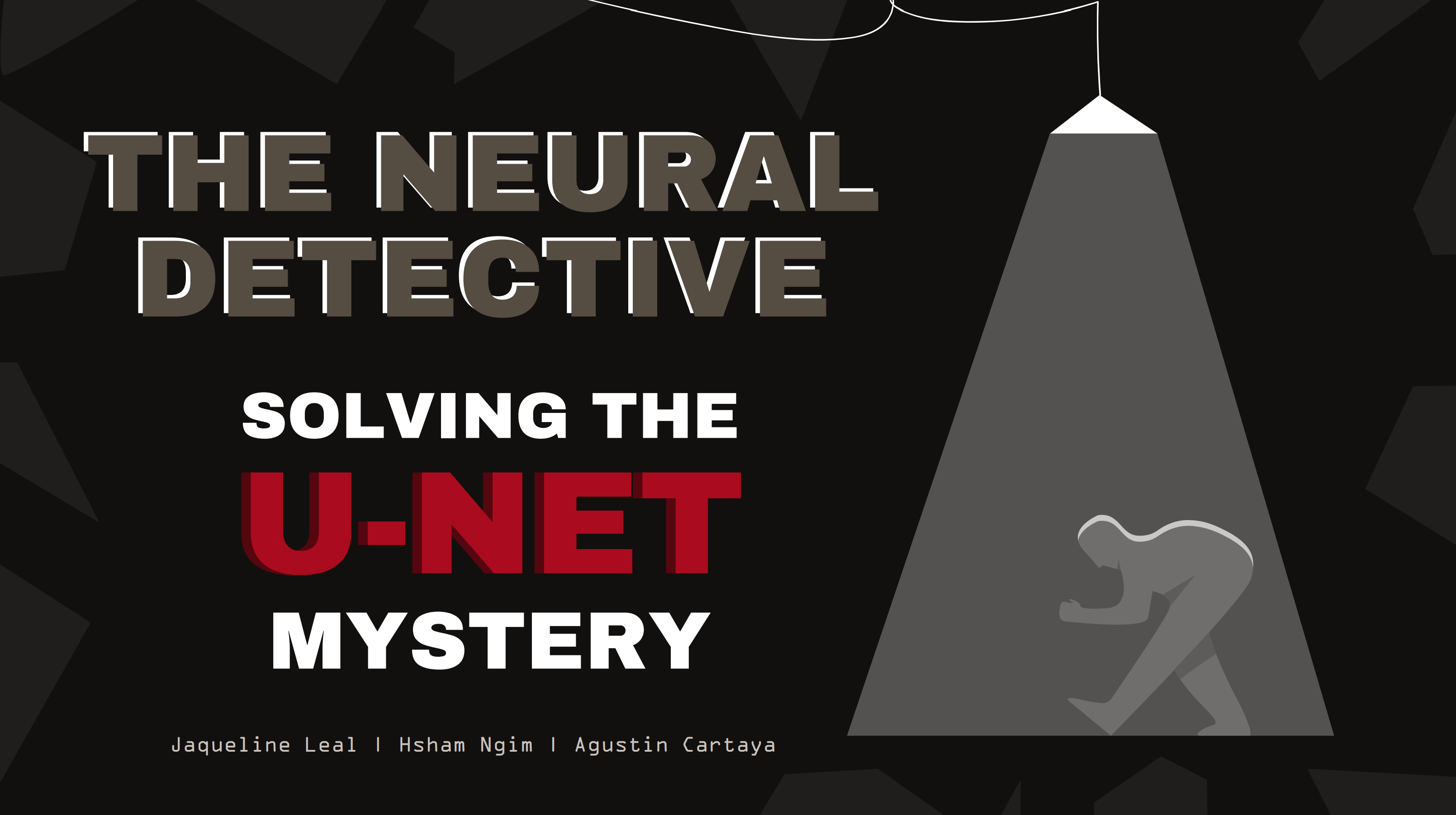
This project proposes an engaging, interactive approach to teach about U-Net's components through a detective-themed game. Participants investigate the causes of poor segmentation results for Dr. Luna, a data scientist, exploring the U-Net architecture to identify key components. This hands-on experience enhances understanding while emphasizing the real-world importance of improving segmentation in medical imaging. This report outlines the steps taken to develop the project and gather participant feedback.
Brain Tumour Detection Using Dempster Shafer Combination
ImViA, Dijon France
September 2023
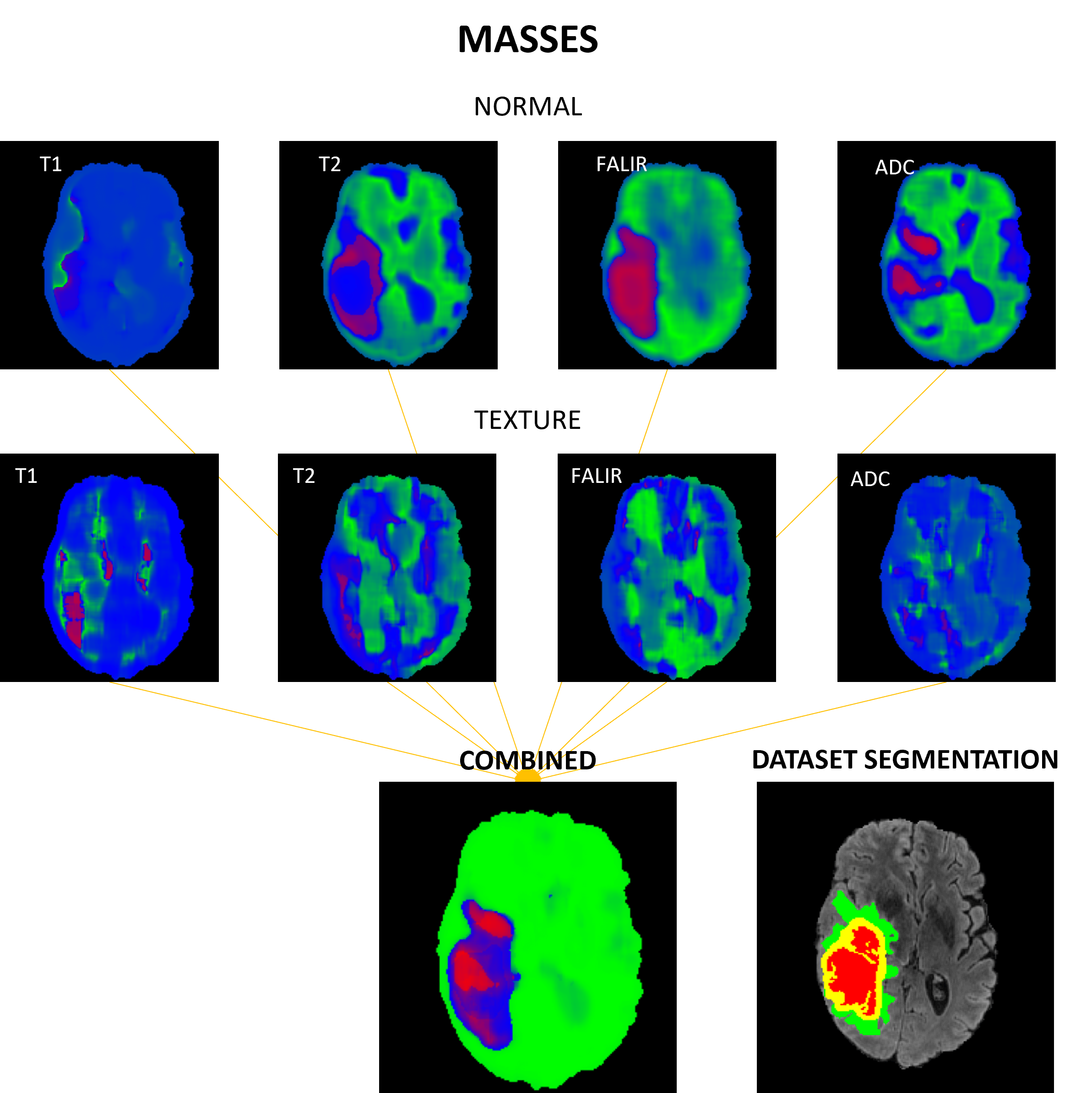
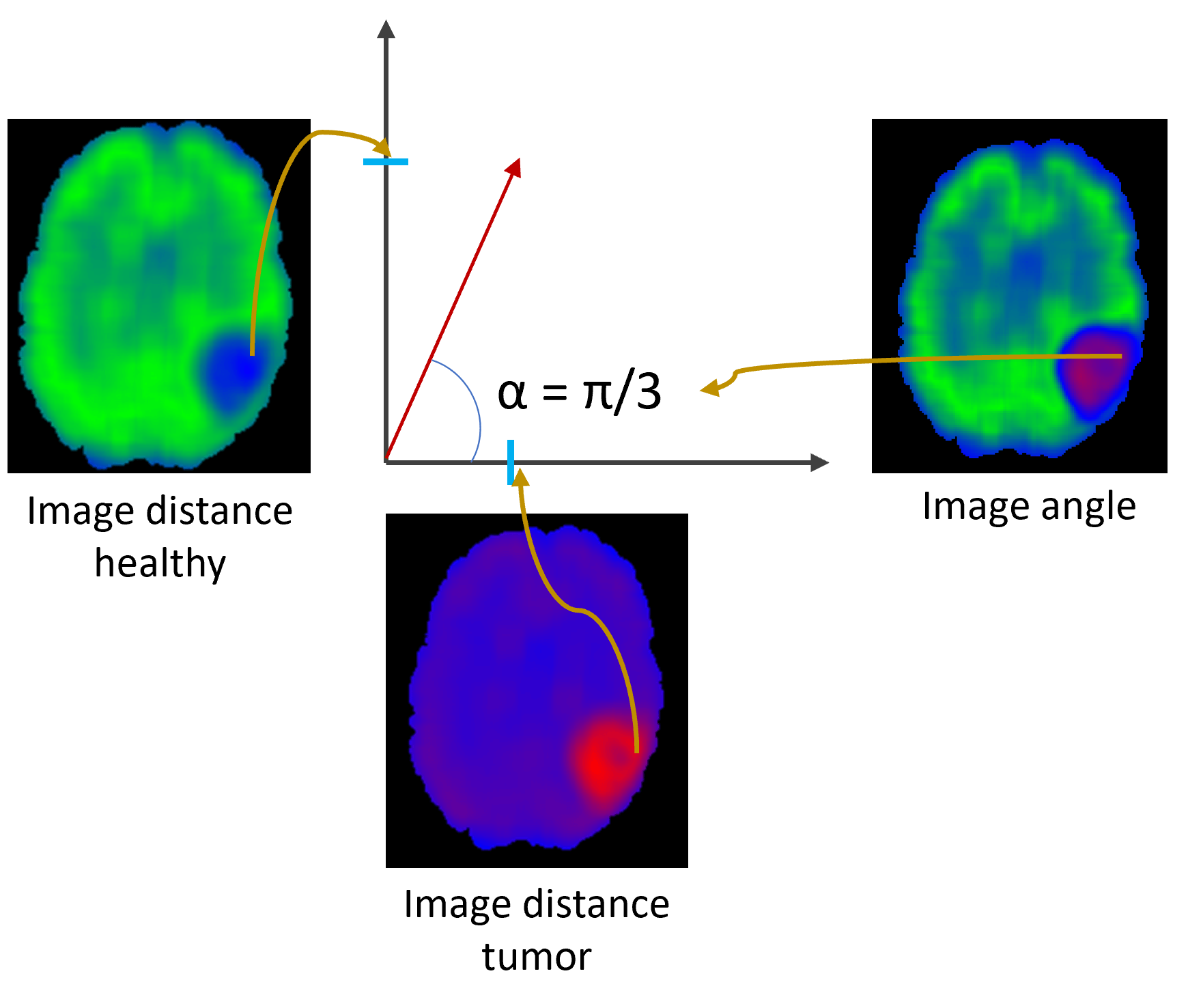
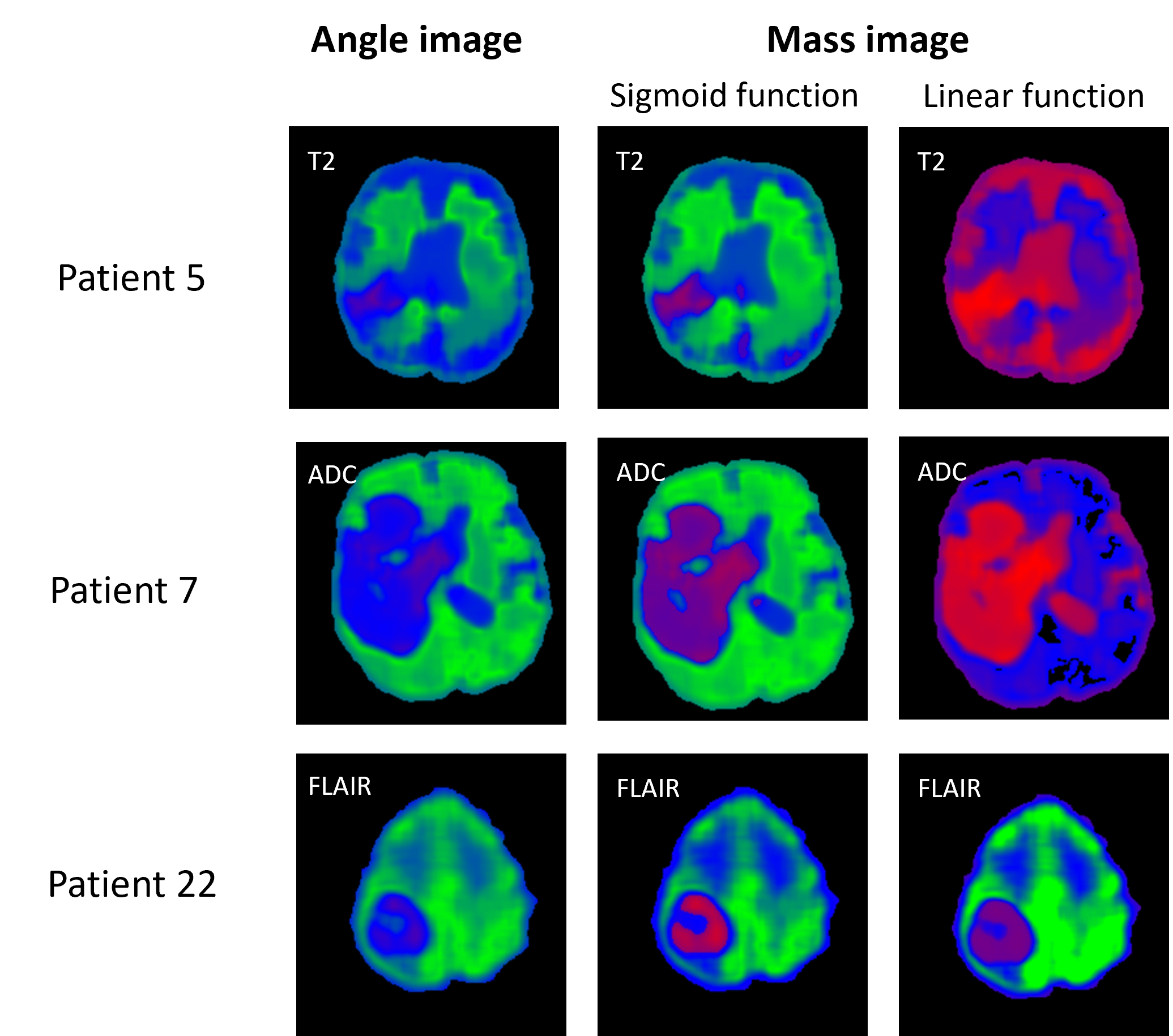
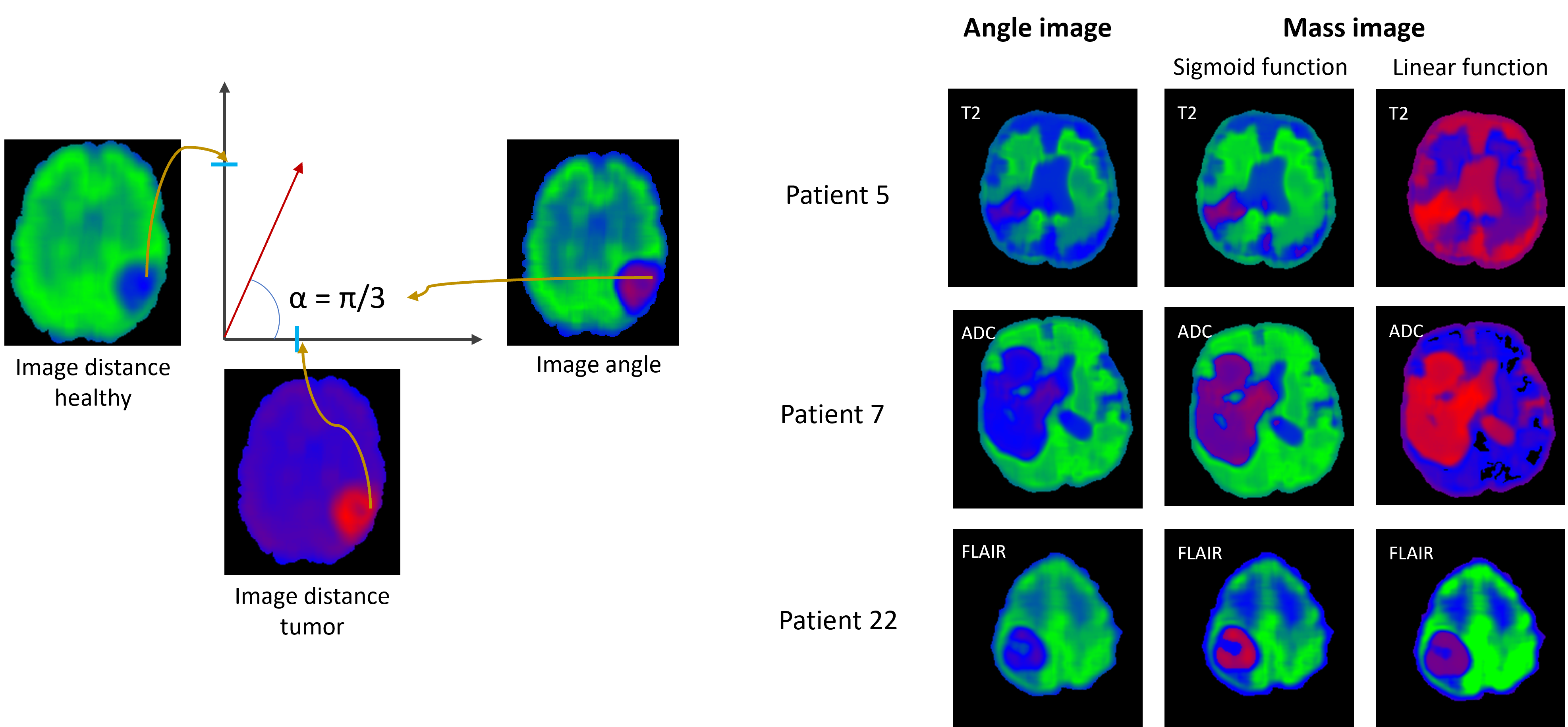
This project focuses on brain tumor segmentation from MRI images. The proposed method integrates data from multiple MRI modalities—T1, T2, FLAIR, and ADC—using the Dempster-Shafer theory to achieve 3D tumor segmentation. In this approach, each modality generates 3D maps where each voxel is assigned a probability between 0 and 1, indicating the likelihood of being part of a tumor. These probabilities are then combined using Dempster’s rule, resulting in a final image with improved certainty about tumor presence. The method was applied to 3D images from glioma patients in the UCSF-PDGM database.
Alzheimer's Disease Classification using MRI and Gene Expression Data
UNICAS, Cassino, Italy
June 2023
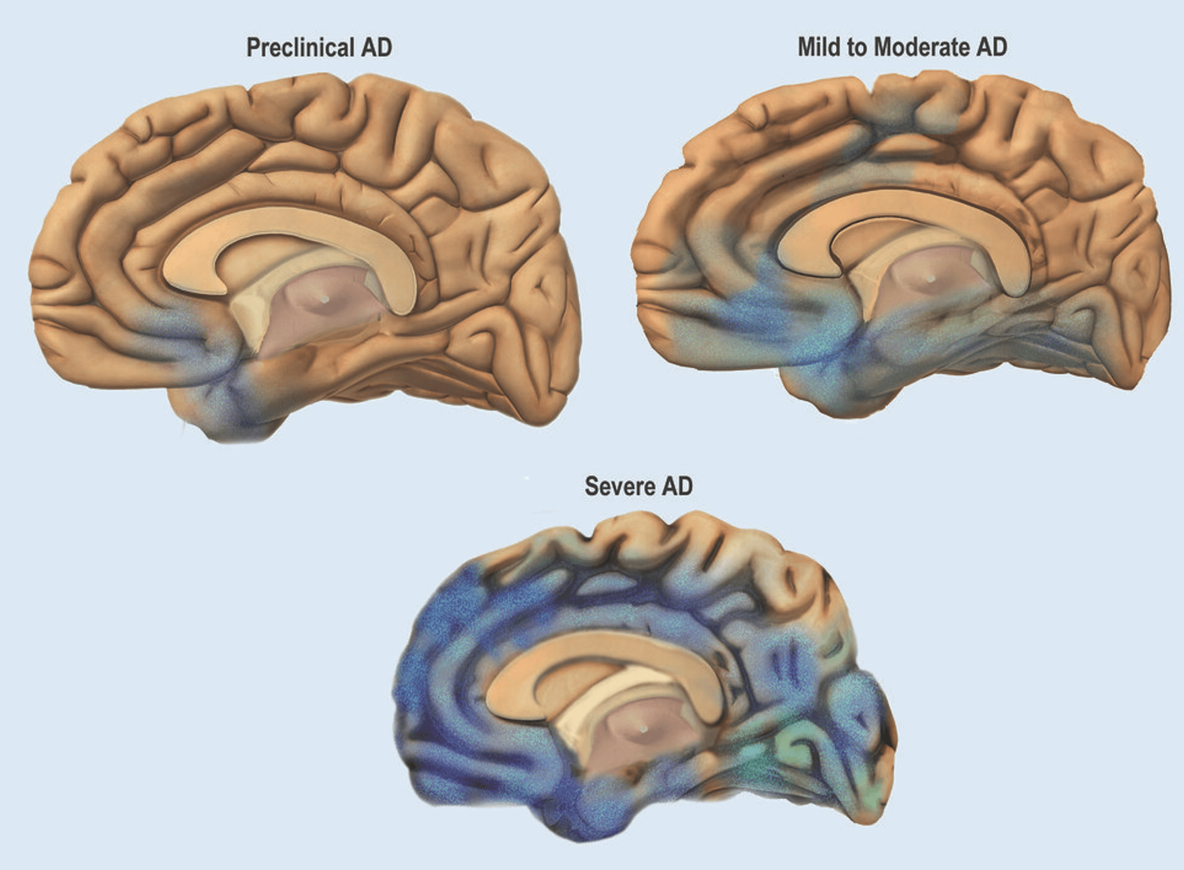
This project focuses on developing machine learning models to diagnose Alzheimer's Disease (AD) and its early stages using MRI scans and gene expression data. We aim to classify patients into different stages of cognitive decline: Alzheimer’s Disease (AD), Mild Cognitive Impairment (MCI), and healthy controls (CTL). The models are trained on a combination of medical imaging and genetic data, using advanced feature selection and classification techniques to optimize prediction accuracy. The goal is to contribute to early diagnosis and improve understanding of Alzheimer’s progression through data-driven analysis.
Alzheimers disease onset recognition by handwriting
UNICAS, Cassino, Italy
June 2023
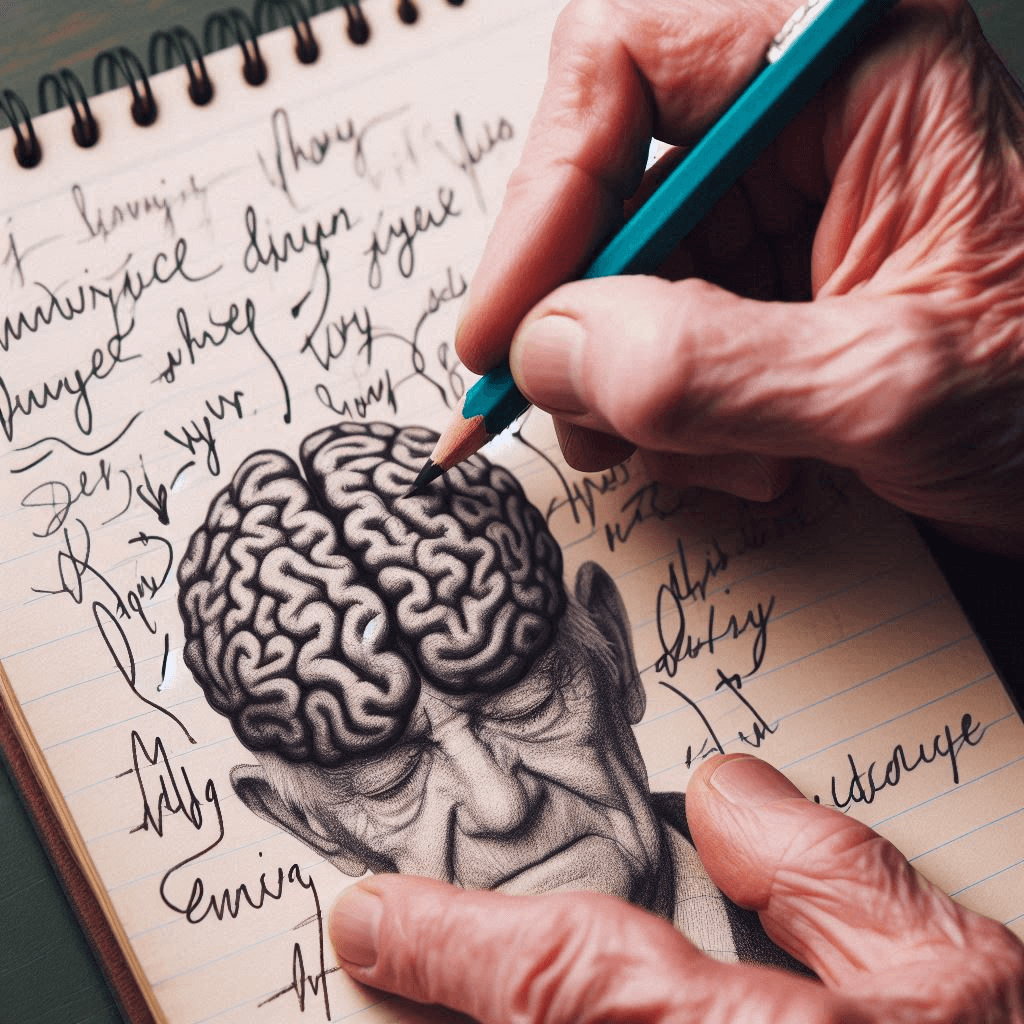
This study combines machine learning (ML) and deep learning (DL) for early Alzheimer’s detection using handwriting analysis. We utilized three approaches: (1) Image analysis with deep learning architectures like VGG19, ResNet50, and Inception to extract deep features from handwriting images, (2) Machine learning classifiers such as Decision Tree, Gradient Boosting, and SVM leveraging deep learning features for enhanced accuracy, and (3) Pen gesture dynamics analysis, examining both on-air and on-paper gestures to identify motor impairments. This multi-faceted approach significantly improves early Alzheimer’s diagnosis by distinguishing between healthy individuals and those with cognitive impairments. The original research project can be found in link
Soft Exudates Detection in Fundus Images
UNICAS, Cassino, Italy
June 2023
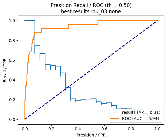
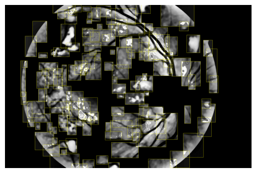
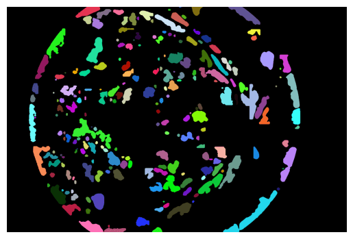
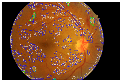
This project introduces a comprehensive approach for the early detection of diabetic retinopathy through the identification of soft exudates (SEx) in fundus images. The process begins with preprocessing to enhance image quality, addressing challenges like variable exudate appearance and low contrast. Additionally, an innovative optical disk removal technique has been implemented to eliminate potential false positives. Then, a two-tiered segmentation strategy is employed: basic segmentation isolates potential SEx areas, while advanced segmentation refines these regions for more precise analysis. Subsequently, feature extraction is performed, extracting key characteristics from the segmented areas to accurately distinguish SEx from other retinal lesions. Finally, sophisticated machine learning algorithms are applied for the classification of SEx.
Skin lesion application – FORTHEM project
ImViA, Dijon France
July 2022
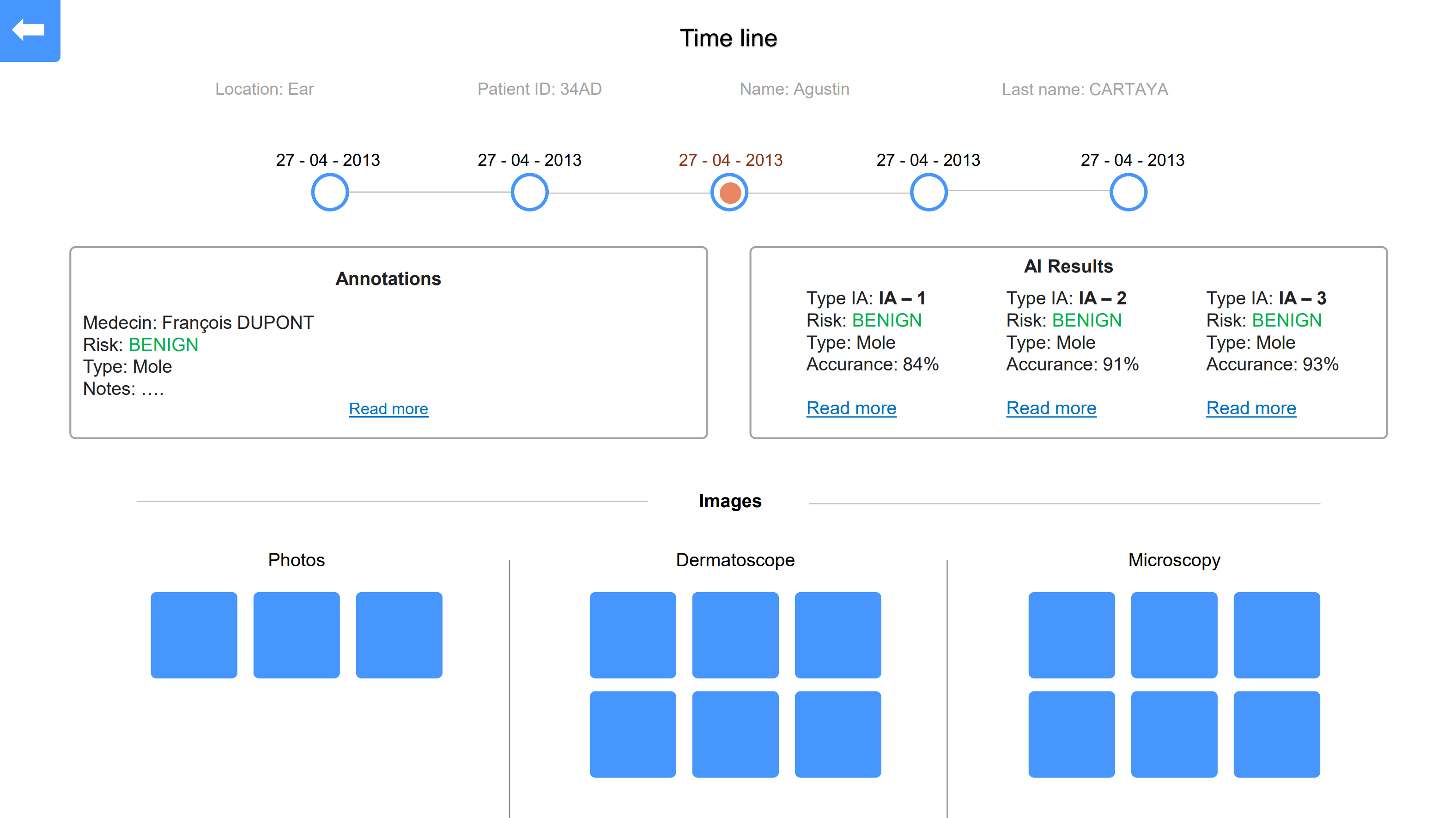
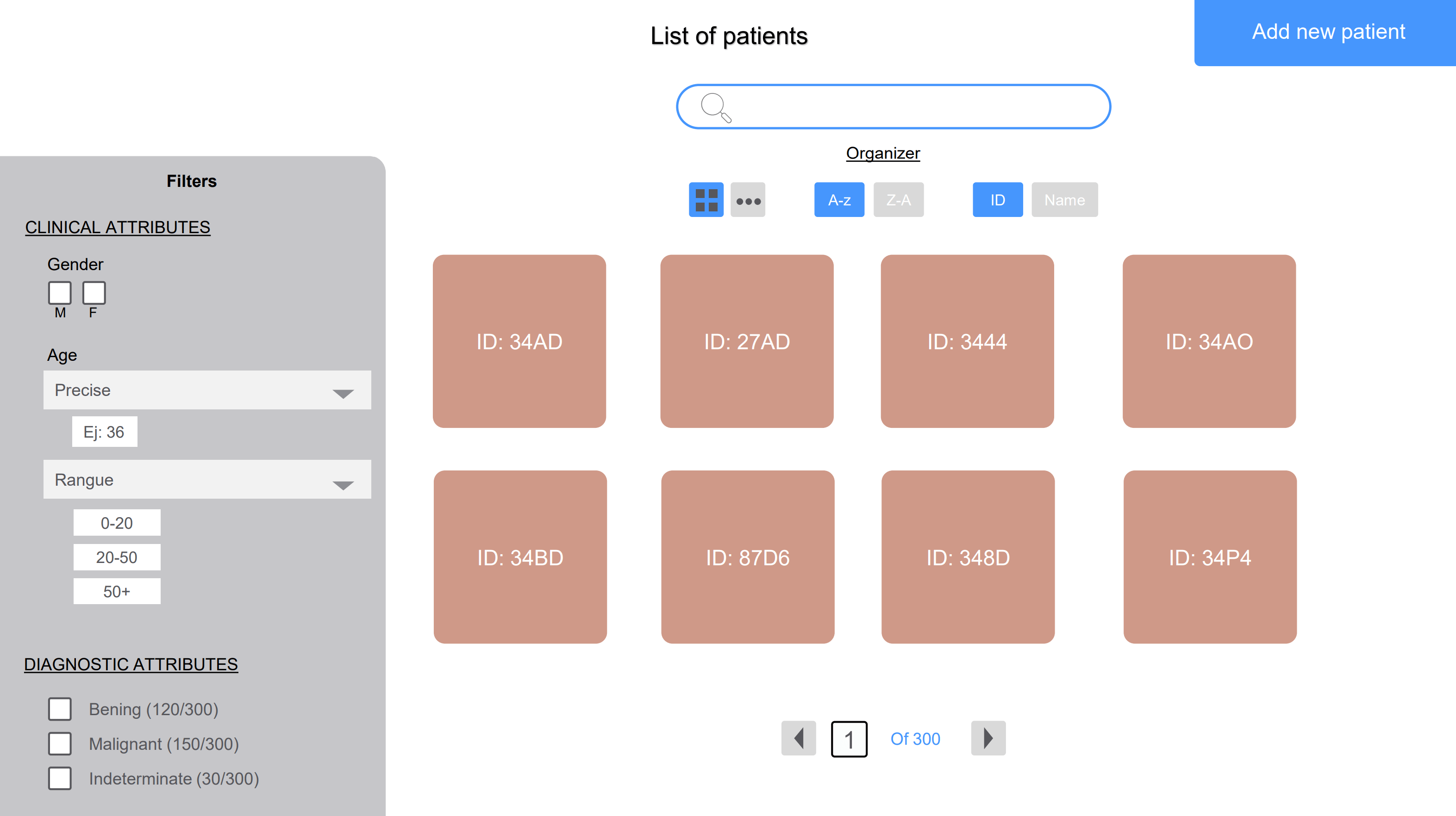
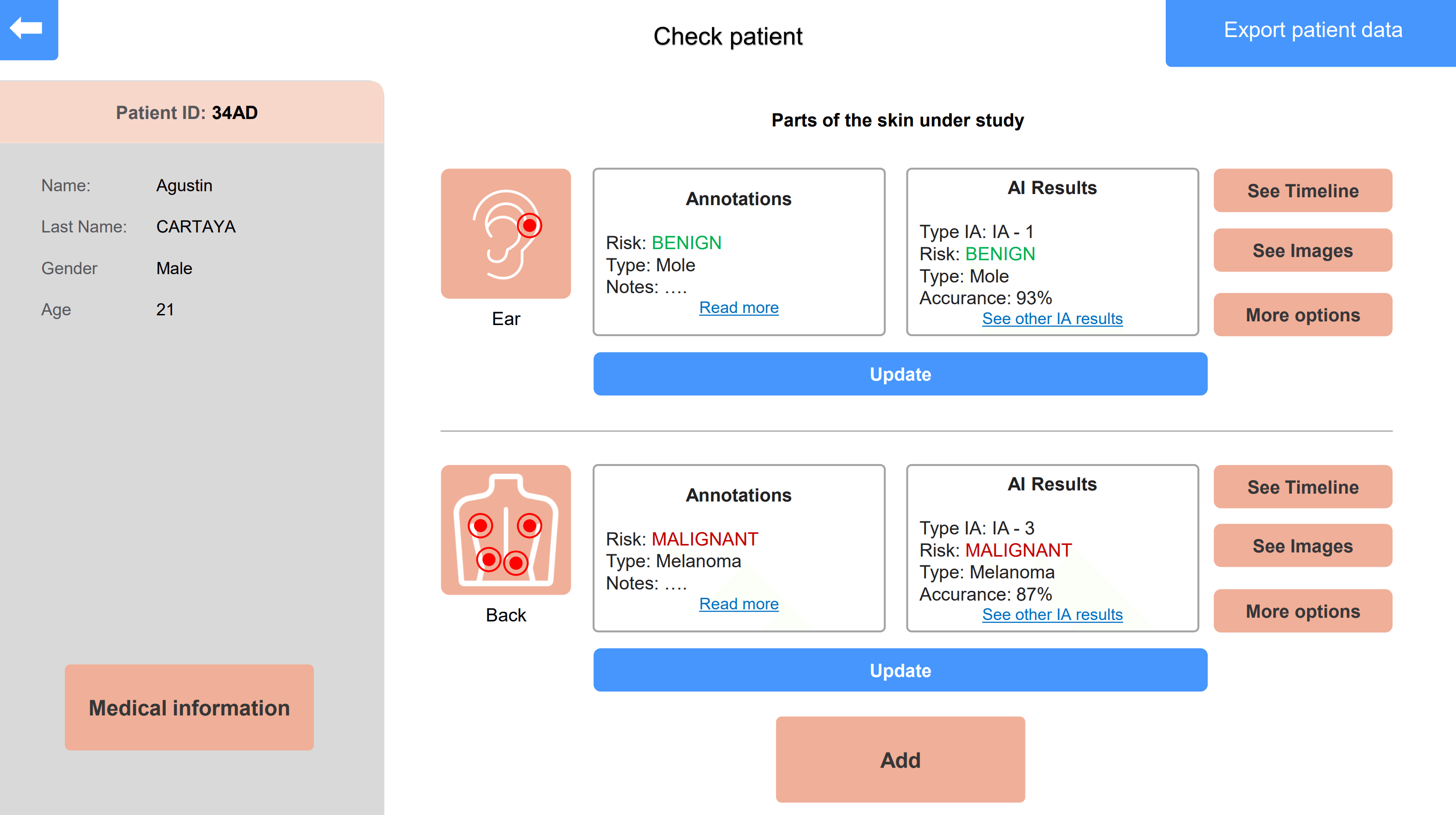
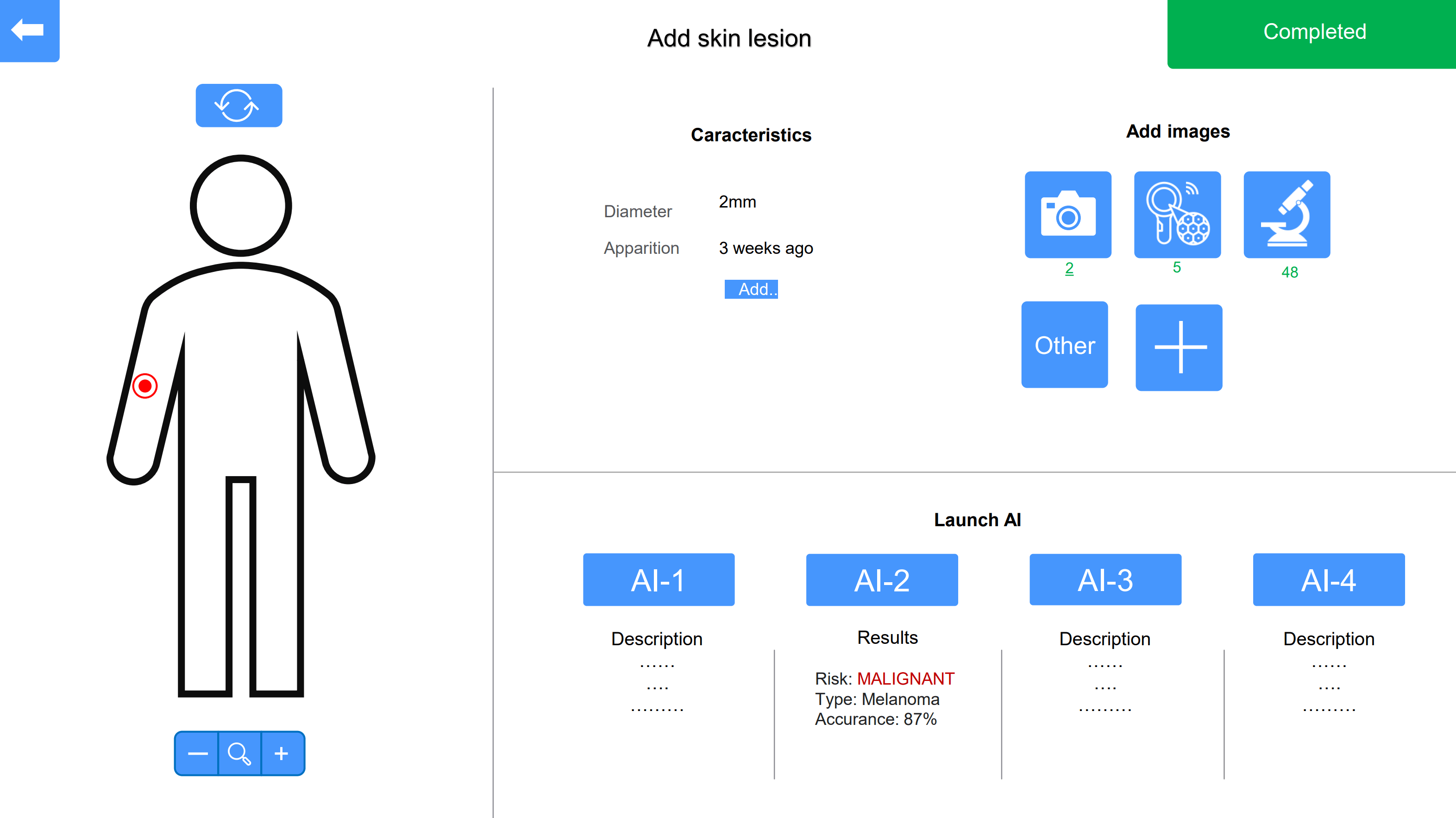
Development of a software application to assist in the analysis of dermatological lesions by integrating multimodal imaging and AI-driven diagnostics. The tool organizes and tracks images from various modalities, such as dermoscopy, reflective confocal microscopy, and photography, providing a comprehensive view of skin lesions. It supports multiple AI systems, utilizing both patient data and lesion characteristics to enhance diagnostic accuracy. Key features include the ability to add new image types, filter patients by lesion data, and visualize lesion progression over time. This software supports dermatologists in managing images and improving diagnostics, particularly in the early detection of skin cancer.
